Urinary Tract Disorders in Cavalier King Charles Spaniels
-
 Xanthinuria
 - • Symptoms
- • Diagnosis
- • DNA Testing
- • Treatment
- Follicular Cystitis
- Research News
- Veterinary Resources
While urinary tract disorders caused by bacterial infections are common among all breeds of dogs, one such disorder, xanthinuria, is much more common in the cavalier King Charles spaniel. A much rarer, inflammatory disease of the bladder, follicular cystitis, has been reported in cavaliers more than in any other breed of dog.
Xanthinuria
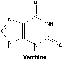
Xanthine uroliths are crystals or sediment in the dog's urinary tract. The disorder, called xanthinuria, is the failure of xanthine, a natural product of the metabolism or purine*, to be dissolved by a process involving xanthine oxidase, and then secreted into the urine. Instead the xanthine concentrates into solids which remain in the urinary tract. The formation of crystals in the urinary tract is called urolithiasis.
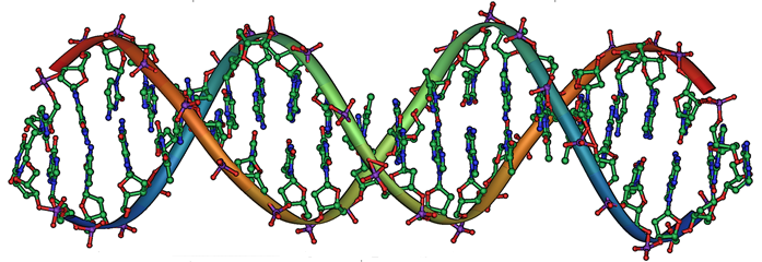
*Purine is an organic compound which have many bodily functions, including serving as building blocks for DNA, aiding blood flow, oxygen delivery, inflammatory responses, and absorption of nutrients.
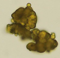 Urolithiasis is a relatively common problem in canines, but
the formation of naturally
occuring xanthine urolit crystals (see photo at left) is very rare. However, it has been found
often enough in related cavalier King Charles spaniels (CKCS) to
conclude that it may be an hereditary defect in the breed, due to
an autosomal (non-sex-linked) recessive mode of inheritance. See this
November 1997 article and this
August 2011 article. But in an
August 2013 article involving 35 CKCSs, they concluded that
"asymptomatic xanthinuria was not detected in this UK Cavalier King
Charles spaniel population."
Urolithiasis is a relatively common problem in canines, but
the formation of naturally
occuring xanthine urolit crystals (see photo at left) is very rare. However, it has been found
often enough in related cavalier King Charles spaniels (CKCS) to
conclude that it may be an hereditary defect in the breed, due to
an autosomal (non-sex-linked) recessive mode of inheritance. See this
November 1997 article and this
August 2011 article. But in an
August 2013 article involving 35 CKCSs, they concluded that
"asymptomatic xanthinuria was not detected in this UK Cavalier King
Charles spaniel population."
RETURN TO TOP
• Symptoms
Cavaliers affected with xanthinuria usually are asymptomatic. Observed symptoms include painful or difficult urination, very slow urination, and a continual or recurrent inclination to attempt to urinate. Also, in some cases, the urine may include some blood. Affected dogs may also display an abnormally great thirst.
RETURN TO TOP
• Diagnosis
In addition to the customary bodily examination and observation of symptoms, diagnosis is determined by blood and urine analyses, urinary untrasounography, and retraction of the stones by urinary cathether and repeated flushing, aspiration, and bladder agitation to retrieve the stones for microscopic analysis. Urine typically would include inflammatory urine sediment, including pyuria (white blood cells), hematuria (red blood cells), and proteinuria (Unusually high amounts of protein).
In the August 2011 article, urolith analysis confirmed pure xanthine. Spot urine samples were submitted for purine analysis along with four controls. Uric acid was detected in all four controls but was not in the CKCS sample. Xanthine and hypoxanthine were detected in the CKCS sample but not in any controls.
RETURN TO TOP
 • DNA
Testing
• DNA
Testing
Research evidence indicates that xanthinuria may be an hereditary defect in the cavalier breed, due to an autosomal (non-sex-linked) recessive mode of inheritance.
In a June 2016 abstract, the researchers reported finding that two cavaliers had an homozygous mutation resulting in a premature stop codon in molybdenum cofactor sulfurase (MOCOS, type 2 xanthinuria). Mutation-specific assays were developed to genotype 108 CKCSs without a history of urolithiasis. Of the 108 cavaliers, 105 were clear, three were carriers, and none were homozygous for the CKCS mutation. They concluded that:
"[D]iverse mutations underlie canine hereditary xanthinuria. Genetic testing can help inform breeders and identify dogs that may benefit from preventative therapies. Future studies are needed to determine mutation frequencies in other breeds and whether clinical outcome differs between the mutations."
The Canine Genetics Laboratory at the University of Minnesota's veterinary school offers an inexpensive genetic test to determine if dogs have the CKCS mutation (or either of two others) causing hereditary xanthinuria. This would be a simple means of confirming whether cavalier breeding stock has the mutation, if the blood line has a history of crystals or sediment in the dogs' urinary tracts.
RETURN TO TOP
• Treatment
The usual treatment for dogs with xanthinuria is to flush the crystals from the body with fluids and to feed them a low-protein alkalizing diet. Eggs, nuts, and dairy products are generally low purine sources of protein. Foods with high purine content (to be avoided) are liver, bacon, kidney, and most seafoods. If flushing is not successful, surgical removal of the stones is necessary.
Allopurinol (Zyloprim) is a drug used to treat dogs suffering from urate bladder stones and from leishmaniasis, a parasite common in certain localities. Allopurinol is an inhibitor of xanthine oxidase, which means that it unfortunately tends to enable the creation of xanthine crystals. In a June 2016 article, researchers studied 320 dogs while receiving allopurinol treatment for leishmaniasis. They found that 29 of the dogs developed xanthinuria and related urinary conditions. Of those dogs, 19 displayed urinary clinical signs. This has been described as "acquired xanthinuria", to distinguish it from the naturally occuring form of xanthinuria in cavaliers. Therefore, care should be taken to not prescribe allopurinol to cavaliers either to treat them for xanthinuria or for leishmaniasis.
A team of ACVIM small animal specialists have issued an ACVIM Consensus Statement on recommnedations for treating and preventing uroliths in dogs (and cats), in a September 2016 article. Regarding xanthinuria (xanthine oxidase), they focus on administering allopurinol resulting in the formation of xanthine uroliths. They explain that it "occurs because allopurinol inhibits the metabolism of xanthine to uric acid and because xanthine is less soluble in urine than is uric acid." They therefore recommend a lower dosage of allopurinol "to safely prevent urate uroliths."
In a November 2018 article, Japanese veterinary researchers report finding that when watermelon extract beverage was substituted for water for 12 dogs for 3 months, the dogs' serum leptin levels were reduced and inhibited the formation of urine crystals such as calcium oxalate and struvite crystals.
RETURN TO TOP
Follicular Cystitis
Follicular cystitis is a rare, inflammatory disease of the bladder which primarily affects females. In a report in August 2014 the first case of follicular cystitis in a dog, a female cavalier King Charles spaniel, was reported. Cystitis is inflammation of the bladder. Most cases of the inflammation are caused by a bacterial infection, and they are called a urinary tract infection (UTI). Follicular cystitis is characterized by the presence of lymphoid follicles with germinal center formation. See also this September 2015 article.
RETURN TO TOP
Research News
November 2018: Watermelon extract found to inhibit formation of urine crystals in dogs. In a November 2018 article, Japanese veterinary researchers (Sayaka Miyai, Toshiharu Hashizume, Toshio Okazaki) report finding that when watermelon extract beverage was substituted for water for 12 dogs for 3 months, the dogs' serum leptin levels were reduced and inhibited the formation of urine crystals such as calcium oxalate and struvite crystals.
August 2017:
University of Minnesota's Canine Genetics Laboratory offers a
genetic test for CKCS xanthinuria.
![]() The
Canine Genetics Laboratory at the University of Minnesota's veterinary school
offers an inexpensive genetic test to determine if dogs have the
CKCS mutation (or either of two others) causing hereditary xanthinuria.
This would be a simple means of confirming whether cavalier breeding
stock has the mutation, if the blood line has a history of crystals or
sediment in the dogs' urinary tracts.
The
Canine Genetics Laboratory at the University of Minnesota's veterinary school
offers an inexpensive genetic test to determine if dogs have the
CKCS mutation (or either of two others) causing hereditary xanthinuria.
This would be a simple means of confirming whether cavalier breeding
stock has the mutation, if the blood line has a history of crystals or
sediment in the dogs' urinary tracts.
September 2016: ACVIM
issues "Consensus Recommendations on the Treatment and Prevention of
Uroliths in Dogs".
 A team of ACVIM small animal specialists
(Jody P. Lulich [right], A.C. Berent, L.G. Adams, J.L. Westropp, J.W. Bartges, C.A.
Osborne) have issued an ACVIM Consensus Statement on recommnedations for
treating and preventing uroliths in dogs (and cats), in a
September 2016 article. Regarding xanthinuria (xanthine oxidase),
they focus on administering allopurinol resulting in the formation of
xanthine uroliths. They explain that it "occurs because allopurinol
inhibits the metabolism of xanthine to uric acid and because xanthine is
less soluble in urine than is uric acid." They therefore recommend a
lower dosage of allopurinol "to safely prevent urate uroliths."
A team of ACVIM small animal specialists
(Jody P. Lulich [right], A.C. Berent, L.G. Adams, J.L. Westropp, J.W. Bartges, C.A.
Osborne) have issued an ACVIM Consensus Statement on recommnedations for
treating and preventing uroliths in dogs (and cats), in a
September 2016 article. Regarding xanthinuria (xanthine oxidase),
they focus on administering allopurinol resulting in the formation of
xanthine uroliths. They explain that it "occurs because allopurinol
inhibits the metabolism of xanthine to uric acid and because xanthine is
less soluble in urine than is uric acid." They therefore recommend a
lower dosage of allopurinol "to safely prevent urate uroliths."
June 2016: University of Minnesota researchers locate a DNA mutation causing hereditary xanthinuria in cavaliers. In a June 2016 abstract, University of Minnesota researchers (Eva Furrow, Nicole Tate, Katie Minor, James Mickelson, Kasey Peterson, Jody Lulich) reported finding that two cavaliers had an homozygous mutation xaresulting in a premature stop codon in molybdenum cofactor sulfurase (MOCOS). Mutation-specific assays were developed to genotype 108 CKCSs without a history of urolithiasis. Of the 108 cavaliers, 105 were clear, three were carriers, and none were homozygous for the CKCS mutation. They concluded that:
"[D]iverse mutations underlie canine hereditary xanthinuria. Genetic testing can help inform breeders and identify dogs that may benefit from preventative therapies. Future studies are needed to determine mutation frequencies in other breeds and whether clinical outcome differs between the mutations."
June 2016: Allopurinol causes xanthinuria in dogs treated for leishmaniasis. In a June 2016 article, Spanish researchers (M. Torres, J. Pastor, X. Roura, M. D. Tabar, Espada, A. Font, J. Balasch, M. Planellas) studied 320 dogs treated with allopurinol for canine leishmaniasis. Forty-two of the 320 dogs -- 13% -- developed xanthinuria.
June 2015:
Researchers emphasize importance of differentiating urine crystals in
cavaliers.
In a
June 2015
study, North Carolina State veterinary school researchers (Kaori
Uchiumi Davis, Carol B. Grindem) reported the importance in determining
the type of urine sediments, particularly in cavalier King Charles
spaniels. In this case, the patient was a male castrated Dalmatian with
a stone and sediment in his urine. The crystals (right) did not dissolve in
acetic acid or hydrochloric acid, but did dissolve in 1 N sodium
hydroxide, ruling out ammonium biurates. Using infrared spectroscopy,
the researchers determined that the spectrum of crystals most closely
matched xanthine. The dog had been treated with allopurinol, which is
used to treat dogs with uric acid crystals. However, allopurinol binds
to and inhibits the action of xanthine oxidase, thereby decreasing
conversion of xanthine to uric acid. Xanthine has low urine solubility,
and therefore xanthinuria may trigger xanthine crystalluria or
urolithiasis. In cavaliers, xanthinuria is hereditary. Since the
solubility results in this case suggest that xanthine crystals may be
significantly different than urate crystals, it therefore is important to
differentiate these crystals before treatment.
August 2014: UK vets find follicular cystititis in a cavalier. A team of veterinarians at the University of Glasgow in the UK report in August 2014 the first case of follicular cystitis in a dog, a female cavalier King Charles spaniel. Cystitis is inflammation of the bladder. Most cases of the inflammation are caused by a bacterial infection, and they are called a urinary tract infection (UTI). Follicular cystitis is characterized by the presence of lymphoid follicles with germinal center formation.
RETURN TO TOP
Veterinary Resources
Xanthine-containing urinary calculi in dogs given allopurinol. Ling GV, Ruby AL, Harrold DR, Johnson DL. JAVMA. 1991;198(11):1935-1940. Quote: Clinical features and laboratory findings were evaluated in 10 dogs that formed xanthine-containing urinary calculi during the period that they were given allopurinol (9 to 38 mg/kg of body weight/d). Duration of allopurinol treatment was 5 weeks to 6 years. Of the 10 dogs, 9 (all Dalmatians) had formed uric acid-containing calculi at least once before allopurinol treatment was initiated. It was not possible to recognize xanthine as a crystalline component of the calculi by use of a chemical colorimetric method or by polarized light microscopy. We concluded that the best diagnostic method for recognition of xanthine-containing calculi was high-pressure liquid chromatography because it is quantitative, sensitive, and accurate, and can be conducted on a small amount (1 to 2 mg) of crystalline material.
Xanthinuria (xanthine oxidase deficiency) in two cavalier king charles spaniels. C.D. van Zuilen , R.F. Nickel, D‐J. Reijngoud. Vet. Qtrly. April 1996;18:sup1-24-25. Quote: A 7-month-old male Cavalier King Charles spaniel was referred to the Department of Clinical Sciences of Companion Animals, Faculty of Veterinary Medicine, Utrecht University, because signs of dysuria and stranguria. ... One littermate and the control dog had normal concentrations of hypoxanthine and xanthine in the urine. The patient and the other littermate had high levels of hypoxanthine and xanthine in the urine (HPLC method and identification witILUV absorption spectrum). Xanthinuria is a well-known but rare hereditary disorder in man (2), characterized by a gross deficiency of the enzyme xanthine oxidase. This enzyme catalyzes the conversion of hypoxanthine to xanthine and of xanthine to uric acid, the end product of purine metabolism in man and higher primates. ... Only two cases of naturally-occurring xanthine calculi in dogs have been reported. One concerned renal calculi in a King Charles spanieland the other concerned urethral uroliths in a Cavalier King Charles spaniel. Neither dog had received any allopurinol medication. Allopurinol is an inhibitor of xanthine oxidase and administration of excessive doses of this drug can cause hyperxanthinuna and formation of xanthine calculi. Neither of the Cavalier King Charles spaniels in this report had received any allopurinol medication. The fact that two littermates were shown to have xanthine oxidase deficiency suggests the possibility of a hereditary xanthine oxidase deficiency in Cavalier King Charles spaniels. (See. also, the November 1997 article below.)
Xanthinuria in a family of Cavalier King Charles Spaniels.
 C.D. van Zuilen, R.F. Nickel, T.H. van Dijk, D‐J. Reijngoud.
Vet.Quarterly. November 1997;19:172-174. Quote: "Xanthine calculi were
found in a 7-month-old male Cavalier King Charles spaniel
with urethral obstruction and renal insufficiency. Because the only two
other reported cases of naturally occurring xanthine urolithiasis
concerned a Cavalier King Charles and a King Charles
spaniel the urine of the littermates and parents of the patient were
also examined for xanthinuria. Semi-quantitative analysis revealed high
urine concentrations of hypoxanthine and xanthine in the patient and his
female littermate. Quantitative analysis by high-pressure liquid
chromatography (HPLC) of the urine samples from the family of this
Cavalier King Charles spaniel and nine control dogs
revealed that hypoxanthine and xanthine excretion was 30 and 60 times
higher in the affected patient and the female littermate than in the
others dogs. The pattern of xanthinuria, which is caused by a deficiency
of the enzyme xanthine oxidase, in the relation diagram of this family
of Cavalier King Charles Spaniels was consistent with
an autosomal recessive mode of inheritance. (See, also, the
April 1996 article above.)
C.D. van Zuilen, R.F. Nickel, T.H. van Dijk, D‐J. Reijngoud.
Vet.Quarterly. November 1997;19:172-174. Quote: "Xanthine calculi were
found in a 7-month-old male Cavalier King Charles spaniel
with urethral obstruction and renal insufficiency. Because the only two
other reported cases of naturally occurring xanthine urolithiasis
concerned a Cavalier King Charles and a King Charles
spaniel the urine of the littermates and parents of the patient were
also examined for xanthinuria. Semi-quantitative analysis revealed high
urine concentrations of hypoxanthine and xanthine in the patient and his
female littermate. Quantitative analysis by high-pressure liquid
chromatography (HPLC) of the urine samples from the family of this
Cavalier King Charles spaniel and nine control dogs
revealed that hypoxanthine and xanthine excretion was 30 and 60 times
higher in the affected patient and the female littermate than in the
others dogs. The pattern of xanthinuria, which is caused by a deficiency
of the enzyme xanthine oxidase, in the relation diagram of this family
of Cavalier King Charles Spaniels was consistent with
an autosomal recessive mode of inheritance. (See, also, the
April 1996 article above.)
Xanthine urolithiasis in a Cavalier King Charles spaniel. Gow, A.G., Fairbanks, L.D., Simpson, J.W., Jacinto, A.M., Ridyard, A.E. Vet. Rec. August 2011;169:209. Quote: "This case is a CKCS in the UK with xanthine urolithiasis and urine purine concentrations after medical management. A four-year old entire male CKCS was referred to the R(D)SVS with a history of polydipsia and intermittent strangury. ... Spot urine samples were submitted for purine analysis along with four controls. Uric acid was detected in all controls but was not in the CKCS sample. Xanthine and hypoxanthine were detected in the CKCS sample but not in any controls. This was suggestive of a defect in purine metabolism, likely a xanthine dehydrogenase deficiency. ... Primary xanthinuria reported in the CKCS is thought to be a genetic defect inherited in an autosomal recessive manner, as in humans). The defect may be a type I or II XDH deficiency. Administration of allopurinol and metabolite analysis would differentiate these; however as this would not alter management, and also have required a control population, this was not performed. XDH activity may also be measured in hepatic or intestinal tissue), this was also not ethically justified. ... Xanthine deposition is thought to cause calculogenic pyelonephritis. This case presented with signs of renal failure which is consistent with the four other symptomatic cases reported. ... In this case, the diet fed was alkalinizing and the urinary pH was 7, the recommended pH for human urine in primary xanthinuria management. In vitro studies and clinical cases have shown minimal dissolution at physiological pH levels therefore at best this is a preventative measure. Although urine specific gravity reduced subsequent to initial presentation, it is unknown whether this was due to improved oral water intake or a deterioration of renal function and poor concentrating ability. In humans, xanthine concentrations below 3 mmol/l are suggested to reduce urolith formation. This was not consistently achieved and uroliths recurred. Urinary xanthine excretion normalised to creatinine appeared to increase compared to the first measurement. This may have been due to poor dietary compliance."
Urine Concentrations of Purine Metabolites in UK Cavalier King Charles Spaniels. .M.L. Jacinto, R.J. Mellanby, M.L. Chandler, N.X. Bommer, H. Carruthers, L.D. Fairbanks, A. Gow. J.Vet.Intern.Med. Nov. 2012; 26(6):1505-1538. Quote: "Xanthine urolithiasis is a rare condition accounting for 0.1% of all canine urolithiasis in one study. This pathology has been reported as a primary disorder in dogs, most notably in Cavalier King Charles Spaniels (CKCS). Xanthine is an intermediate product of purine metabolism, which is converted from hypoxanthine by xanthine oxidase. Xanthine is only slightly soluble in urine and therefore hyperxanthinuria may lead to urolith formation. It has been speculated that some CKCS have an inherited mutation in the xanthine oxidase gene. In humans, isolated deficiency of xanthine oxidase occurs rarely and approximately 50% of individuals are asymptomatic, despite having significant xanthinuria. Therefore we hypothesised that asymptomatic xanthinuria may be commonplace in the UK population of CKCS. In support of this, a previous case report of a symptomatic CKCS reported significant xanthinuria occurring in an asymptomatic sibling. In order to examine the prevalence of xanthinuria in CKCS, urine concentrations of hypoxanthine and xanthine metabolites as well as creatinine were measured in 35 client-owned Cavalier King Charles Spaniel dogs and 24 dogs of other breeds from three first-opinion veterinary practices in the UK. Urine samples were collected by free catch and purine metabolites were measured by high-performance liquid chromatography. Ratios of xanthine/creatinine and hypoxanthine/creatinine from the two populations were compared by Mann Whitney U test and were found not to be significantly different (p=0.41 and p=0.59 respectively). In the control population, the xanthine/creatinine ratio ranged from 0.00018 to 0.01611 (median 0.00069), while in the CKCS population it ranged from 0.000154 to 0.005794 (median 0.000435). These results are markedly lower than the previously reported case of xanthine urolithiasis in a UK CKCS dog, which utilised the same reference laboratory (xanthine/creatinine ratio 0.406). These data suggest that asymptomatic xanthinuria is not prevalent in the UK CKCS population."
Urine concentrations of xanthine, hypoxanthine and uric acid in UK Cavalier King Charles spaniels. Ana Margarida Leça Jacinto, Marjorie Chandler, Nick X. Bommer, Harvey Carruthers, Lynette D. Fairbanks, Richard J. Mellanby, Adam G. Gow. J. Small Anim. Pract. August 2013;54(8):395-398. Quote: "Objectives: Xanthine urolithiasis and asymptomatic xanthinuria have been diagnosed in Cavalier King Charles spaniel dogs suggesting that primary xanthinuria may be a breed-related disorder, although its prevalence remains unclear. The hypothesis of this study was that asymptomatic xanthinuria is common in Cavalier King Charles spaniel dogs. Methods: Free catch urine samples were collected from 35 client-owned Cavalier King Charles spaniel dogs and from 24 dogs of other breeds. The purine metabolites were measured by high-performance liquid chromatography. The urine ratios of xanthine/ creatinine and hypoxanthine/creatinine were calculated and compared between the two groups of dogs. Results: The urine concentrations of purine metabolites were not significantly different between the two groups and were very low in both. The urine concentrations of xanthine in all 35 Cavalier King Charles spaniel were markedly lower than in the previously reported case of xanthine urolithiasis in a UK Cavalier King Charles spaniel dog. Clinical Significance: Asymptomatic xanthinuria was not detected in this UK Cavalier King Charles spaniel population. This data may be used as a reference for urinary purine metabolite concentrations in the dog."
Follicular cystitis in a dog. Rui Moncao Sul, Gawain Hammond, Kathryn Pratschke. Vet.Rec. Case Report. August 2014;2(1). Quote: A four-year-old female Cavalier King Charles Spaniel with a history of recurrent lower urinary tract disease refractory to treatment was referred to our hospital. Clinical examination was unremarkable apart from thickening of the dorsal vulva. Abdominal ultrasound was compatible with possible areas of mild thickening of the bladder wall. Lower genitourinary-contrast radiographic studies showed multiple small lesions in the bladder wall. Surgical biopsy of the bladder was compatible with follicular cystitis and excised uterine tissue was consistent with cervicitis. Clinical signs resolved after treatment with a combination of antibiosis, NSAIDs, pentosan polysulfate and amitriptyline. Follow-up 30 months after surgery confirmed that the dog was free of clinical signs. Follicular cystitis has not been previously reported in dogs but should be considered as a differential for patients with refractory long-standing lower urinary tract disease.
What is your diagnosis? Urine crystals from a dog. Kaori Uchiumi Davis, Carol B. Grindem. Vet. Clinical Pathology. June 2015;44(2):331-332. Quote: "A 5-year-old male castrated Dalmatian weighing 19.5 kg was evaluated by the referring veterinarian for being unable to urinate. A lateral radiograph showed potential mineralized material in the ureters. The dog was administered buprenorphine [Temgesic sublingual] and acepromazine ... Additional radiographs were taken at North Carolina State University Veterinary Health Complex (NCSU-VHC) which did not reveal a radiopaque stone. Urinary catheterization was performed and a stone was found in the urethra and flushed into the bladder. The dog was prescribed allopurinol 300 mg BID and the stranguria resolved. The dog was reevaluated 2 months later. A urinalysis with sediment examination was performed and revealed mild alkalinuria (pH 9.0; reference interval 4.5-8.5). Urine was opaque golden colored with a specific gravity of 1.023, pH of 9, trace protein, negative glucose, trace ketone, and 1 + bilirubin. A sulfosalicylic acid protein precipitation test was trace positive. Examination of sediment revealed rare epithelial cells, many fat droplets, and many yellow-brown small spherules of ammonium biurate-like crystals. The crystals did not dissolve in acetic acid or hydrochloric acid, but did dissolve in 1 N sodium hydroxide, ruling out ammonium biurates. The spectrum of crystals determined by infrared spectroscopy most closely matched xanthine. Xanthine is a product of purine metabolism and is converted to uric acid by the enzyme xanthine oxidase. Xanthine crystals are impossible to distinguish from ammonium biurate or amorphous urates by light microscopy. All of these crystals have yellow-brown color and appear globular to amorphous. Infrared spectroscopy or high-pressure liquid chromatography can be used to confirm xanthine crystalluria. Xanthine crystalluria and urolithiasis are rare in small animals; uroliths with at least 70% xanthine were reported to account for less than 0.1% of all canine uroliths. It usually occurs secondary to therapy with allopurinol. Xanthinuria associated with allopurinol therapy is influenced by several variables including the dosage of allopurinol, quantity of dietary purine precursors, the rate of degradation, and hepatic function. Allopurinol is used to treat dogs with uric acid crystals. It is also used to treat infectious diseases such as leishmaniasis and trypanosomiasis, as it is incorporated into RNA, leading to blockage of de novo synthesis of purine nucleotides. It binds to and inhibits the action of xanthine oxidase, thereby decreasing conversion of xanthine to uric acid. Xanthine has low urine solubility, and therefore xanthinuria may trigger xanthine crystalluria or urolithiasis. Besides allopurinol, differential diagnoses for xanthinuria include hereditary xanthinuria (Cavalier King Charles Spaniels) or excessive dietary purines. Prevention involves low purine intake, high water intake, and urine alkalinization to reduce urine xanthine concentration and increased its solubility. It is unclear why the dog had highly alkaline urine. It could be due to alkalinization in an attempt to dissolve the stone, or a false increase in pH due to contamination of cleaning solution. Xanthine urolithiasis has been reported in cats, dogs, and people. Information on solubility of xanthine crystals in the literature is lacking, but solubility results in the present case suggest that they may be significantly different than urate crystals and important in differentiating these crystals."
Ureteral implantation using a three-stitch ureteroneocystostomy: description of technique and outcome in nine dogs. K. M. Pratschke. J. Sm. Anim. Pract. September 2015;56(9):566-571. Quote: Objective: To report the procedure, postoperative outcome and complications of a new technique for ureteral implantation by means of a three-stitch ureteroneocystostomy in dogs. Materials & Methods: Clinical records of dogs requiring ureteral implantation between April 2007 and June 2013 were retrospectively reviewed. Data retrieved included signalment, preoperative biochemistry results, details of the surgical procedure, perioperative and postoperative complications, postoperative biochemistry results and outcome. Results: Nine dogs [including a female neutered cavalier King Charles spaniel with left ureteral obstruction by suspected inflammatory granuloma and concurrent follicular cystitis] fulfilled the inclusion criteria. Follow-up times ranged from 10 to 79 months (median 30 months), with 8 of 9 dogs having an excellent long-term outcome and no major postoperative complications. One dog with follicular cystitis as a comorbidity developed obstruction from inflammatory granuloma and required revision surgery. Clinical Significance: The three-stitch technique for ureteral implantation compares favourably to previously documented techniques in terms of outcome and complication rates. Reduced tissue handling and a decreased volume of suture material may be beneficial for healing. The technique is also faster than previously described options, which may be of benefit in unstable patients requiring ureteral implantation due to traumatic injury or rupture.
Adverse urinary effects of allopurinol in dogs with leishmaniasis. M. Torres, J. Pastor, X. Roura, M. D. Tabar, Espada, A. Font, J. Balasch, M. Planellas. J. Sm. Anim. Pract. June 2016;57(6):299-304. Quote: Objective: The objective of this study was to describe the adverse effects of allopurinol on the urinary system during treatment of canine leishmaniasis. Methods: Retrospective case series of 42 dogs that developed xanthinuria while receiving allopurinol treatment for leishmaniasis. Results: Of 320 dogs diagnosed with leishmaniasis, 42 (13%) developed adverse urinary effects. Thirteen (of 42) dogs (31%) developed xanthinuria, renal mineralisation and urolithiasis; 11 (26·2%) showed xanthinuria with renal mineralisation; 9 (21·4%) had xanthinuria with urolithiasis and 9 (21·4%) developed xanthinuria alone. Urinary clinical signs developed in 19 dogs (45·2%). Clinical Significance: This study demonstrates that urolithiasis and renal mineralisation can occur in dogs receiving allopurinol therapy. Dogs receiving therapy should be monitored for the development of urinary adverse effects from the beginning of treatment.
Three Diverse Mutations Underlying Canine Xanthine Urolithiasis. Eva Furrow, Nicole Tate, Katie Minor, James Mickelson, Kasey Peterson, Jody Lulich. J. Vet. Int. Med. June 2016. 2016 ACVIM Forum Research Report Program. Quote: Hereditary xanthinuria in people is an autosomal recessive disease caused by mutations in xanthine dehydrogenase (XDH) or molybdenum cofactor sulfurase (MOCOS). There are rare reports of hereditary xanthinuria in dogs, but genetic investigations have not previously been described. The objective of this study was to identify mutations underlying risk for canine xanthine urolithiasis by sequencing XDH and MOCOS in affected dogs. Five dogs with primary xanthine urolithiasis (i.e. no history of xanthine dehydrogenase inhibitor therapy) were recruited. This group included 2 Cavalier King Charles Spaniels (CKCS), 2 Toy Manchester Terriers (TMT), and a mixed-breed dog. Three homozygous mutations were identified. The CKCSs had a mutation resulting in a premature stop codon in MOCOS, the TMTs had a splice site mutation in MOCOS, and the mixed breed dog had a splice site mutation in XDH. cDNA sequencing confirmed exon skipping for the splice site mutations. Mutation-specific assays were developed to genotype 108 CKCSs and 50 TMTs without a history of urolithiasis. Of the 108 CKCS, 105 were clear, 3 were carriers, and none were homozygous for the CKCS mutation. Of the 49 TMTs, 37 were clear, 10 were carriers, and 2 were homozygous for the TMT mutation. Urine was analyzed from the 2 homozygous TMTs and revealed xanthinuria. In conclusion, diverse mutations underlie canine hereditary xanthinuria. Genetic testing can help inform breeders and identify dogs that may benefit from preventative therapies. Future studies are needed to determine mutation frequencies in other breeds and whether clinical outcome differs between the mutations. See, also: Three diverse mutations underlying canine xanthine urolithiasis. J. Animal Sci. September 2016;94(7supp4):163(P6030). N. M. Tate, K. M. Minor, J. R. Mickelson, K. Peterson, J. P. Lulich, E. Furrow. Quote: Hereditary xanthinuria in people is an autosomal recessive disease caused by mutations in xanthine dehydrogenase (XDH) or molybdenum cofactor sulfurase (MOCOS). There are rare reports of hereditary xanthinuria in dogs, but genetic investigations have not previously been described. The purpose of this study was to uncover mutations underlying risk for canine xanthine urolithiasis by sequencing XDH and MOCOS in genomic DNA from affected dogs. The affected dogs included two Toy Manchester Terriers (TMT), two Cavalier King Charles Spaniels (CKCS), and a mixed breed dog. Three putative causal mutations were found. The TMT dogs had a homozygous splice site mutation in MOCOS, the CKCS dogs had a homozygous nonsense mutation in MOCOS, and the mixed breed dog had a homozygous splice site mutation in XDH. cDNA sequencing verified aberrant splicing for the two splice site mutations. Mutation assays were developed to determine the allele frequencies of the mutations in populations of TMT and CKCS dogs without a history of xanthine urolithiasis. Of 49 TMT dogs tested, 37 were clear, 10 were carriers, and 2 were homozygous for the TMT mutation. Urine was analyzed from the 2 homozygous TMTs and revealed xanthinuria. Of 108 CKCS dogs tested, 105 were clear, 3 were carriers, and none were homozygous for the CKCS mutation. In conclusion, diverse mutations were found to be responsible for hereditary xanthinuria in dogs, and we have developed genetic tests for these forms of the disease. Genetic testing can help inform breeders and identify dogs that may benefit from preventative therapies.
ACVIM Small Animal Consensus Recommendations on the Treatment and Prevention of Uroliths in Dogs and Cats. J.P. Lulich, A.C. Berent, L.G. Adams, J.L. Westropp, J.W. Bartges, C.A. Osborne. J. Vet. Int. Med. September 2016;30(5):1564-1574. Quote: In an age of advancing endoscopic and lithotripsy technologies, the management of urolithiasis poses a unique opportunity to advance compassionate veterinary care, not only for patients with urolithiasis but for those with other urinary diseases as well. The following are consensus-derived, research and experience-supported, patient-centered recommendations for the treatment and prevention of uroliths in dogs and cats utilizing contemporary strategies. ... Recommendation 3.4.a: Consider Xanthine Oxidase Inhibitors for Dogs Homozygous for Genetic Hyperuricosuria that have Failed Therapeutic Diet Prevention: Rationale: Urate urolith recurrence is common, especially in dogs with a genetic mutation in the urate transporter. Prevention may require more than dietary adjustments. The dosage of allopurinol to sufficiently prevent urate urolith recurrence without xanthine urolith formation is variable and influenced by the severity of disease, endogenous purine production, quantity of purines in the diet, urine pH, and urine volume. In a case series of 10 dogs with previous urate urolithiasis, allopurinol administration in excess of 9-38 mg/kg/d was associated with xanthine urolith formation. This occurs because allopurinol inhibits the metabolism of xanthine to uric acid and because xanthine is less soluble in urine than is uric acid. Based on these observations, we recommend a dosage of 5-7 mg/kg q12-24 h to safely prevent urate uroliths. The role and effectiveness of allopurinol and newer-generation xanthine oxidase inhibitors in patients with porto-vascular shunts are unknown. Administration of xanthine oxidase inhibitors should be avoided in dogs that are not receiving decreased purine diets to minimize the risk of xanthine urolith formation. ... Ultimately, we hope that these recommendations will serve as a foundation for ongoing and future clinical research and inspiration for innovative problem solving.
Effects of a Watermelon Extract Beverage on Canine Lipid Metabolism and Urine Crystals. Sayaka Miyai, Toshiharu Hashizume, Toshio Okazaki. Anim. & Vet. Sci. November 2018;6(5):74-79. Quote: Previous report showed that watermelon consumption has an anti-obesity effects in rats. The purpose of this study is to examine the effects on body weight, body fat percentage, serum biochemical data, serum adipokine concentrations (leptin, adiponectin, and resistin), urine specific gravity and sediments when 12 dogs were given watermelon extract beverage instead of water for 3 months. Those data were all assessed before the study period and again at 1.5 and 3 months. Although there were no remarkable changes in most of these parameters, a significant decrease in serum leptin concentrations at 1.5 and 3 months. Calcium oxalate and struvite crystals were observed in the urinary sediment in five dogs; although their urine specific gravities remained >1.040 throughout, the number of urinary crystals had decreased by the end of the 3-month period. Morphological components were not found in the urinary sediment of the other five dogs; their urine specific gravities were also >1.040 before the study period and at 1.5 months, but these had decreased to <1.040 at 3 months. These results suggested that drinking the watermelon extract beverage reduced serum leptin levels and inhibited the formation of urine crystals such as calcium oxalate and struvite crystals in dogs.
Urolithiasis in Small Animals. Alice Defarges, Michelle Evason, Marilyn Dunn, Allyson Berent. Clinical Small Animal Internal Medicine, Chap. 123. April 2020; doi: 10.1002/9781119501237.ch123. Quote: Formation of urinary calculi (uroliths) has been hypothesized to occur through multiple mechanisms or processes. Three of the more common of these theories are the precipitation‐crystallization theory, the matrix‐nucleation theory, and the crystallization‐inhibition theory. Urine is commonly supersaturated with crystalloids, and this is a prerequisite for urolith formation. Factors that predispose to urine stasis also play an important role in urolithiasis formation. Risk factors for calcium oxalate uroliths in dogs include sex, and they occur more commonly in male than female dogs. ... Xanthine is an important mediator in purine metabolism; the drug allopurinol binds rapidly to (and inhibits the action of) xanthine oxidase, thereby decreasing conversion of hypoxanthine to xanthine and xanthine to uric acid. The result is a reduction of serum and urine concentrations of uric acid, with an increase in serum and urine concentrations of xanthine. As such, most xanthine uroliths in dogs form secondary to therapy with allopurinol via this mechanism. This is particularly true when a diet high in purines (meat based) is fed to these at‐risk dogs. Naturally occurring xanthinuria has been reported in Cavalier King Charles spaniels and dachshunds, and is thought to reflect an inborn error of xanthine oxidase activity. ... Hepatic dysfunction is associated with a reduced ability to convert ammonia to urea and uric acid to allantoin. Therefore, dogs suffering from hepatic dysfunction may develop hyperammonuria and hyperuricuria, which may result in urate urolith formation. Urolithiasis typically induces inflammatory urine sediment, such as pyuria (presence of white blood cells), hematuria (red blood cells), and proteinuria.
CONNECT WITH US